
Ongoing research projects
Our overarching research interest is to investigate the multifaceted role of imaging in the full spectrum of autoimmune rheumatic and degenerative musculoskeletal diseases. Ongoing research projects are dedicated to understanding the role of inflammation and structural damage in joint tenderness in different arthritides, the anatomic basis of inflammation in small joints of the hands, as well as utility of novel PET tracers in the diagnosis and prediction of interstitial lung disease in connective tissue diseases, such as myositis
Tendon involvement in osteoarthritis of the hand
Tenosynovitis is an inflammatory process affecting the synovial sheath of tendons which occurs in various rheumatic and musculoskeletal diseases. It is associated with pain and persistent tenosynovitis, and can lead to adhesions and dysfunction. However, data on tendon involvement in hand osteoarthritis, a highly prevalent musculoskeletal condition, is limited. The aim of this study, performed within the framework of the Ludwig Boltzman Institute for Arthritis and Rehabilitation was to characterize tendon involvement in HOA, assess its frequency and influence on pain and hand function, as well as its potential association with the presence of osteophytes. The project is financed by the Mayor´s Medical-Scientific Fund of Vienna.
OMERACT Ultrasound Working Group
Members of our research group are actively involved in multiple subgroups of the OMERACT Ultrasound Working Group and have contributed to or led the development of sonographic outcome measures in RMDs, including cartilage change and structural damage in rheumatoid arthritis, sonographic lesions in gout and calcium-pyrophosphate deposition, enthesitis, dactylitis and synovitis. Current work features standardizing the consensus finding process and reliability exercises, which are the hallmark of the group.

Development of outcome instruments based on imaging according to the OMERACT stepwise model, showing the four steps of the selection and development process. Terslev L et al. J Rheum. 2019;46:1394-1400.
FAPI Imaging
Interstitial lung disease and heart involvement are common manifestations or comorbidities of rheumatic and musculoskeletal diseases in general and connective tissue diseases in particular, and a substantial contributor to hospitalisation, increased morbidity and mortality. In collaboration with the Division of Nuclear Medicine MUW, we are investigating the utility of ⁶⁸Ga-labelled inhibitor of Fibroblast-Activation-Protein (FAPi) based positronic emission tomography and computed tomography imaging for visualizing and quantifying fibroblast activation in the lung, heart and muscle of patients with connective tissue disease and other rheumatic and musculoskeletal diseases.

[68Ga]68Ga-DATA5m.SA.FAPi uptake in the lung. Representative computed tomography (A) and [68Ga]68Ga-DATA5m.SA.FAPi PET-CT image of a scan from a patient with interstitial lung disease in idiopathic inflammatory myopathy.
Aptamers in fibroblasts
Efficient, timely treatment of inflammatory arthritis is severely hampered by the limited efficacy of current medications as well as by the lack of definitive markers of disease activity. Aptamers are single-stranded RNA segments that can be selected to bind various target molecules, including proteins and are advantageous over classical antibodies in many aspects. In this joint project with the research group of Tamás Mészáros at the Department of Molecular Biology, Institute of Biochemistry and Molecular Biology of Semmelweis University, Budapest, Hungary, our aim is to generate highly selective aptamers which can neutralize proteins that are known to play a role in the activation of fibroblasts, cells which play a key role in the inflammation of the synovial membrane. Aptamer development yield agents with therapeutic implications, while those developed against as of yet unknown markers of fibroblast activation may aid in characterizing the localization of such cells in joints. The project is financed by the Austrian Science Fund.
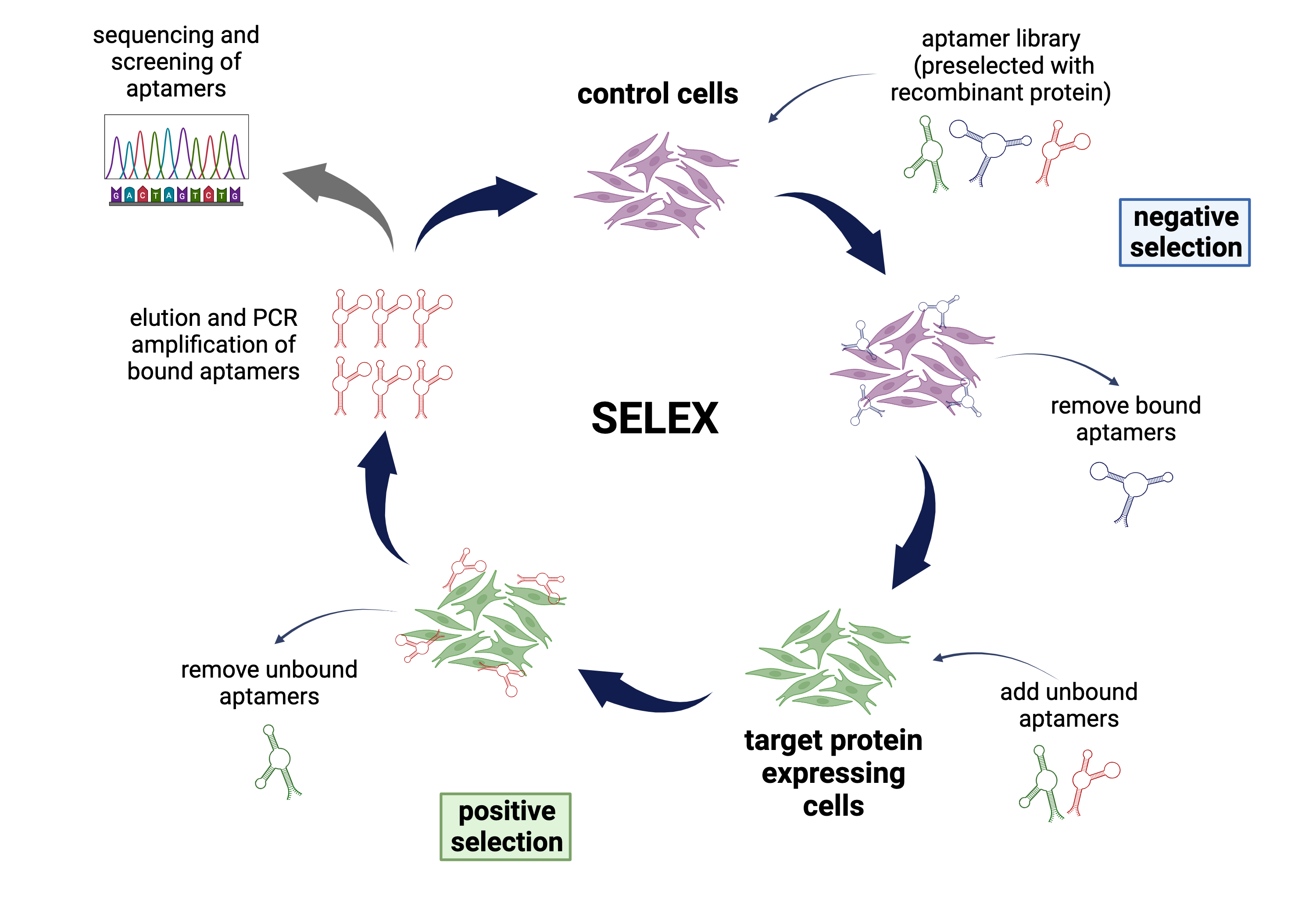
Aptamer selection workflow according to the SELEX (Systematic Evolution of Ligands by EXponential enrichment) process. Design by Jessica Lange.
Anatomy of the image
In collaboration with the University of Barcelona and the Department of Radiology at the MUW we are conducting a comparative imaging study using ultrasound, magnetic resonance imaging and histology exploring the joint capsule of the metacarpophalangeal joints, the contribution of muscles and ligaments to the capsule and the presence and significance of functional and anatomical enthesis within and around the joint.
Tenderness and psychiatric comorbidities
Previous studies led by Irina Gessl in our group have explored whether joint tenderness can be considered an objective or rather a subjective sign of joint inflammation in arthritis. A bidirectional relationship exists between rheumatoid arthritis, psoriatic arthritis and mood and anxiety disorders, which may also influence outcomes in inflammatory arthritis. In addition, subjective illness perception was also reported to be associated with patient reported outcomes longitudinally independently of inflammation. The goal of this project is to assess the association of depression, anxiety and illness perception with subjective and objective signs of joint inflammation in patients with RA, assess a clustering of the patients according to these variables and ultimately assess a potential benefit of treating/targeting depression, anxiety and illness perception regarding subjective and objective markers of disease activity.
Technology
With regard to clinical research, the Division is equipped with several high-end musculoskeletal ultrasound units and facilities required to perform synovial biopsies. Close collaboration with the Department of Radiology and the Division of Nuclear Medicine ensure access to high-end, cutting-edge imaging techniques and methods. Experimental strategies in the wet lab include state-of-the-art technologies, including modern molecular biology and advanced cell biology techniques, a variety of immunological tools, biochemical and molecular approaches. The use of human patient material as well as in vivo models provides the opportunity to emphasize the translational aspects in all projects performed.
Training
Appropriate training is highly important to ensure skilled use and accurate interpretation of findings by rheumatologists. There is a growing network of European centres performing research on imaging in rheumatic diseases. We offer training in novel imaging techniques, in particular musculoskeletal ultrasound as well as in imaging-guided procedures, such as synovial biopsies. Postdocs and PhD students will work on cutting-edge research projects in a collaborative research environment. Interested candidates can participate in the PhD programs of the Medical University of Vienna.
www.meduniwien.ac.at/hp/phd-immunologie/
https://www.meduniwien.ac.at/hp/n790-mdr/
Existing mentoring programs at the Medical University of Vienna as well as national and international collaborations provide an excellent basis for an internationally successful scientific career in academia and/or the life science industry. Additional training in writing publications and grant applications, reviewing manuscripts and presenting at national and international conferences enables young researchers to build an individual career as an independent researcher.